Picture this: a patient sits in your exam chair with a dense nuclear cataract that has completely blocked your optical biometer. No axial length reading. No reliable IOL calculation. You reach for the DGH A ultrasound, connect the lightweight probe, and within seconds you have consistent, high-quality measurements that let you move forward confidently with surgery planning. That reliable backup is exactly why clinics across the country trust the DGH A every day.
Eye care teams know that precise biometry drives successful cataract outcomes. When optical methods fall short, ophthalmic ultrasound steps in to deliver the axial length measurements, anterior chamber depth, and lens thickness data you need for accurate intraocular lens power calculation. The DGH A makes this process straightforward, portable, and clinically precise.
Understanding Ocular Biometry and the Role of DGH A Ultrasound
Ocular biometry measures the eye’s key dimensions to calculate the perfect IOL power before cataract surgery. Axial length measurement sits at the heart of every formula, and even a 0.1 mm error can shift refraction by nearly a diopter. The DGH A ultrasound uses a 10 MHz ultrasonic transducer to send sound waves through the eye and capture the echoes from the cornea, lens surfaces, and retina.
Unlike optical devices that rely on light, the DGH A works beautifully through dense cataracts, small pupils, or corneal irregularities. Its pattern recognition software instantly grades each scan with a clear three-star system, so you know right away whether the measurement meets your standards. Clinics love that it also supports myopia management by tracking axial length progression over time with easy-to-read reports.
Applanation Versus Immersion: Choosing the Right Technique with DGH A
You have two practical options with the DGH A, and each fits different clinical situations.
Contact (applanation) mode keeps things quick and simple for routine cases. You gently touch the probe to the anesthetized cornea while the patient fixates on the probe’s red light. The device’s compression lockout helps you avoid artificially short readings.
Immersion mode, using the included Prager shell, eliminates any chance of corneal compression. You fill the shell with sterile saline, rest it on the sclera, and take measurements with the probe suspended in the fluid bath. Many surgeons prefer immersion for premium IOL patients because it delivers the tightest repeatability, often within ±0.03 mm.
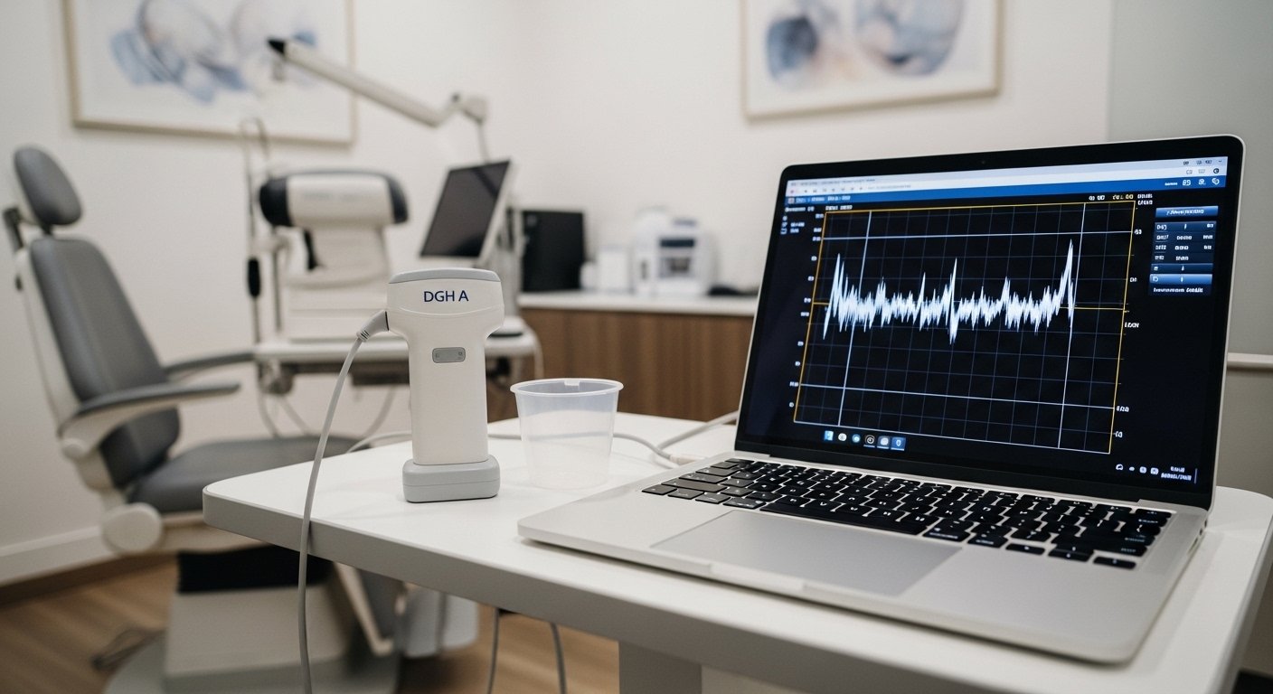
Both techniques produce excellent results when you follow proper alignment. The DGH A’s audible feedback and real-time waveform display guide your hand so you consistently capture high-quality data.
Standout Features of the Portable DGH A Ultrasound Device
The DGH A, also known as the Scanmate 6000, weighs less than a pound and plugs directly into any Windows computer via USB. No bulky console, no extra cart. You simply install the intuitive Scanmate software, enter patient details, and start scanning.
Key advantages include:
- Automatic gain control that optimizes every waveform
- Multiple built-in IOL formulas plus post-refractive options
- EMR compatibility for seamless record keeping
- Axial length progression reports for myopia patients
- Manual measurement review if you ever need to fine-tune a borderline scan
These features make the DGH A a practical choice for busy clinics that want professional-grade biometry without complicated setup.
DGH A Scan Accuracy: How It Compares to Optical Biometry
Optical biometry remains the first choice for clear media because it is non-contact and highly reproducible. Yet studies and daily practice show that the DGH A matches optical results closely when immersion technique is used. In eyes with dense cataracts or other media opacities, the DGH A often provides the only usable data.
The device’s corneal compression detection and pattern recognition help reduce operator variability. Experienced technicians routinely achieve repeatability that supports confident IOL selection. When optical and ultrasonic readings differ, many surgeons average the two or rely on the immersion ultrasound as the tiebreaker. The bottom line: the DGH A does not replace optical biometry, but it completes it perfectly for the 8 to 17 percent of cases where light-based systems fail.
Practical Guide: How to Use DGH A for IOL Power Calculation in Cataract Surgery
Using the DGH A feels straightforward once you run through it a couple of times. Here is the typical workflow:
- Open the Scanmate software and create or select the patient record.
- Choose contact or immersion mode and verify velocities for the patient’s lens status.
- Apply topical anesthetic (and saline for immersion).
- Position the probe, instruct the patient to fixate, and acquire five to ten consistent scans.
- Let the software automatically select the best measurements or manually review the waveforms.
- Switch to the IOL calculator, select your preferred formula, and compare results for different lens models.
- Save the report and export to your EMR.
The entire process usually takes just a few minutes, freeing you to focus on surgical planning rather than troubleshooting equipment.
Essential Maintenance: Cleaning DGH A Scan Probes and Ensuring Longevity
Proper care keeps your DGH A performing like new and protects patients from infection risk. After every use, disconnect the probe and wipe the tip, housing, and cord with a soft cloth dampened in mild soap and water or 70 percent isopropyl alcohol. Follow with an approved disinfectant according to the manufacturer’s guidelines. Never immerse the probe beyond the strain relief area, and always dry it thoroughly before storage.
The immersion shell receives the same cleaning routine. A quick daily inspection of the cable and USB connection prevents unexpected downtime. Most clinics find that simple habits like these keep the device reliable for years.
Integrating DGH A into Your Clinic: Workflow and Software Benefits
The DGH A fits neatly into modern workflows because it runs on the same computer you already use for charting. Patient data flows directly into your database, and reports print or export with one click. Whether you run a high-volume surgical center or a smaller practice, the portability means you can move the unit between lanes without hassle.
Teams especially appreciate the software’s personalized lens constants feature. After a few dozen cases, you can refine your constants for even tighter refractive outcomes. The result is smoother surgery days and happier patients.
Actionable Takeaways You Can Use Tomorrow
- Schedule a quick in-service with your technicians on immersion technique to boost measurement consistency.
- Review your last ten dense-cataract cases and note where ultrasound would have helped.
- Add DGH A cleaning to your end-of-day checklist so the probe stays ready.
- Explore the myopia management reports if you see young patients with progressing axial length.
Three things to try this week: run a side-by-side contact-versus-immersion comparison on a volunteer eye, export a sample report to your EMR, and discuss with your team which IOL formulas you use most often in the calculator.
What has been your experience when optical biometry falls short? Many clinics discover that adding the DGH A removes that stress entirely and gives them confidence on every case.
You May Also Like: Seekde Explained: The New Era of Intent-Driven AI Search
FAQs
How much does a DGH A scan cost for clinics?
The DGH A ultrasound device typically falls in an affordable range that makes it accessible for most modern practices. Contact DGH Technology or authorized distributors for current pricing and any available trade-in options.
How accurate is the DGH A compared to optical biometry?
In clear media, optical devices lead slightly. For dense cataracts or challenging eyes, the DGH A with immersion technique delivers excellent repeatability (often ±0.03 mm) and serves as the trusted backup that keeps your surgical plan on track.
What is the difference between applanation and immersion with DGH A?
Applanation (contact) is faster for routine cases, while immersion uses a saline-filled shell to avoid any corneal compression. Most surgeons choose immersion for premium lenses or when maximum precision matters.
How do you clean and maintain the DGH A scan probes?
Wipe the probe after each use with mild soap and water or 70 percent isopropyl alcohol, followed by approved disinfectant. Dry completely and never submerge past the strain relief. The same simple steps apply to the immersion shell.
Does the DGH A support post-refractive IOL calculations?
Yes. The Scanmate software includes popular post-refractive formulas so you can confidently calculate lens power for patients who previously had LASIK, PRK, or RK.
Is the DGH A compatible with my clinic’s software or EMR?
The device works with standard Windows PCs and offers direct EMR export, making integration smooth for most practices.
How portable is the DGH A ultrasound device?
Extremely. The probe and interface weigh less than a pound total and connect via USB, so you can move it between exam rooms or even take it to satellite clinics without hassle.